Takotsubo syndrome

Takotsubo syndrome is a condition characterized by the sudden onset of acute, transient (lasting up to 3 weeks) dysfunction of the left ventricle’s systolic function. This dysfunction typically occurs in response to intense emotional or physical stress
Hence, Takotsubo syndrome is commonly referred to as ‘broken heart syndrome’ due to its association with emotional stress, though it can also be precipitated by physical stressors
Prevalence
The prevalence of Takotsubo syndrome is estimated to be approximately 2-4% among all patients and 5-6% among women hospitalized with a diagnosis of acute coronary syndrome.
This syndrome predominantly affects women over 50 years of age. This demographic trend may be attributed to heightened sympathetic nervous system activity and diminished parasympathetic nervous system stimuli. Increased sympathetic activity enhances myocardial sensitivity to catecholamines.
Etiology
While the precise cause of Takotsubo syndrome remains elusive, research has substantiated the significant role of psycho-emotional stress in its onset.
Pathogenesis and pathophysiology
Several hypotheses attempt to elucidate the pathogenesis of Takotsubo syndrome.
Adrenergic theory
The theory of endogenous adrenergic surge stands as one of the most extensively researched. In a majority of patients, emotional or physical stress acts as the precipitating factor for Takotsubo syndrome.
Stress triggers the release of significant quantities of catecholamines, which subsequently interact with beta-adrenoceptors. Studies in mice with hyperactive sympathetic nervous systems have revealed a notable increase in beta-adrenoceptor density at the apex of the heart. Some researchers suggest that this surge in adrenoceptor stimulation disrupts myocardial calcium regulation, ultimately leading to left ventricular contractility suppression.
Brain-Heart Axis
Observational data from Takotsubo syndrome patients frequently indicate psychological stress as a significant trigger for the condition. Furthermore, many patients exhibit comorbid mental health disorders such as depression, anxiety, and schizophrenia.
This correlation finds a scientific basis in the altered expression of miRNA 16 and miRNA 26a in patients with depression and anxiety. Overexpression of these miRNAs in rodent models has been shown to induce abnormalities in the movement of the apical segment of the left ventricle.
Neurocardiogenic cardiac stunning, a well-recognized complication following acute neurological injury, manifests as transient systolic dysfunction of the left ventricle in 20-30% of patients. This underscores the intricate interplay between the brain and the heart.
Coronary vasospasm and microvascular reactivity
Microvascular dysfunction and impaired reactivity are hallmark features of Takotsubo syndrome, possibly contributing to its predilection for postmenopausal women. Estrogen, under physiological conditions, enhances coronary blood flow through both endothelium-dependent and -independent mechanisms. Its deficiency can lead to endothelial dysfunction, potentially exacerbating microvascular disturbances observed in Takotsubo syndrome.
Metabolic and energetic disorders
Current evidence indicates the presence of metabolic and energetic abnormalities in Takotsubo syndrome patients. Preclinical studies suggest increased myocardial glucose uptake, accompanied by heightened activity of glycolytic enzymes. Paradoxically, this is coupled with reduced availability of glycolytic metabolites, indicative of disrupted metabolic homeostasis.
Inflammatory mechanisms
Emerging evidence supports the involvement of inflammation in Takotsubo syndrome. However, the role of myocardial inflammation during the acute phase remains multifaceted, potentially serving as both a cause and a consequence of the syndrome. Histological studies have revealed myocardial macrophage infiltration, alongside elevated systemic levels of proinflammatory cytokines in Takotsubo syndrome patients.
Clinical presentation
Most Takotsubo syndrome patients present with acute chest pain and shortness of breath, often precipitated by psychoemotional stress.
In severe cases, Takotsubo syndrome can lead to profound heart failure symptoms, life-threatening arrhythmias, and thromboembolic events.
Medical history
During the collection of anamnesis, it is necessary to find out:
- Whether the patient has a history of mental health disorders such as depression, anxiety syndromes, or schizophrenia, these conditions are recognized risk factors for Takotsubo syndrome.
- Whether the patient has a history of somatic diseases such as diabetes, bronchial asthma, chronic obstructive pulmonary disease (COPD), encephalitis, meningitis, or encephalopathy, these conditions are also associated with an increased risk of Takotsubo syndrome.
- Whether the patient has undergone recent invasive diagnostic or treatment procedures, which can potentially trigger Takotsubo syndrome.
- Whether the patient has recently experienced excessive physical exertion, can be a precipitating factor for Takotsubo syndrome.
- Whether the patient is currently taking medications that stimulate the sympathetic nervous system, such as beta-2 adrenergic receptor agonists, alpha-1 adrenergic receptor agonists, serotonin and norepinephrine reuptake inhibitors (SNRIs), or epinephrine.
- Whether the patient uses psychotropic substances such as cannabis, cannabinoids, or amphetamines, may increase the risk of Takotsubo syndrome.
Physical examination
The physical presentation of Takotsubo syndrome varies depending on the severity of the condition.
In patients with an uncomplicated course of Takotsubo syndrome, a moderate increase in heart rate may be noted.
In patients experiencing complications of Takotsubo syndrome, signs of acute heart failure may be evident. These can include moist wheezing in the lungs, tachypnea, and decreased blood oxygen saturation. Upon auscultation of the heart, tachycardia, an additional pathological third heart sound, and a systolic murmur over the apex of the heart due to obstruction of the left ventricular outflow tract and mitral regurgitation may be present. Additionally, patients with Takotsubo syndrome may exhibit decreased blood pressure.
Assessment of probability
The probability of Takotsubo syndrome is evaluated in patients presenting with chest pain and/or shortness of breath who have been hospitalized in intensive care units, particularly when there is no ST segment elevation observed on the electrocardiogram (ECG).
The InterTAK (The International Takotsubo Diagnostic Criteria) diagnostic scale is employed to assess the likelihood of Takotsubo syndrome, utilizing the following criteria and corresponding points:
| Criteria | Points |
| Female sex | 25 |
| Emotional trigger | 24 |
| Physical trigger | 13 |
| Absence of ST segment depression (except AVR) | 12 |
| Psychiatric disorders | 11 |
| Neurological disorders | 9 |
| Prolongation of the corrected QT interval (women > 460 ms, men > 440 ms) | 6 |
A score exceeding 70 points on this scale indicates a high probability of Takotsubo syndrome (approximately 90%), while a score of 70 points or lower suggests low or moderate probability.
In cases where Takotsubo syndrome is deemed highly probable, urgent transthoracic echocardiography is recommended. Conversely, when the probability is low or moderate, coronary angiography is advised to definitively differentiate Takotsubo syndrome from acute myocardial infarction.
Examination methods
Laboratory examination methods for suspected Takotsubo syndrome include:
- Blood test for troponin I/T;
- Blood test for creatine phosphokinase (CK);
- Blood test for brain natriuretic peptide (BNP) or N-terminal pro-brain natriuretic peptide (NT-proBNP)
Instrumental examination methods for suspected Takotsubo syndrome include:
- Electrocardiography;
- Transthoracic echocardiography;
- Coronary angiography;
- Ventriculography;
- Cardiac magnetic resonance imaging (MRI)
Blood test for troponin I / T and Creatine Phosphokinase (CK)
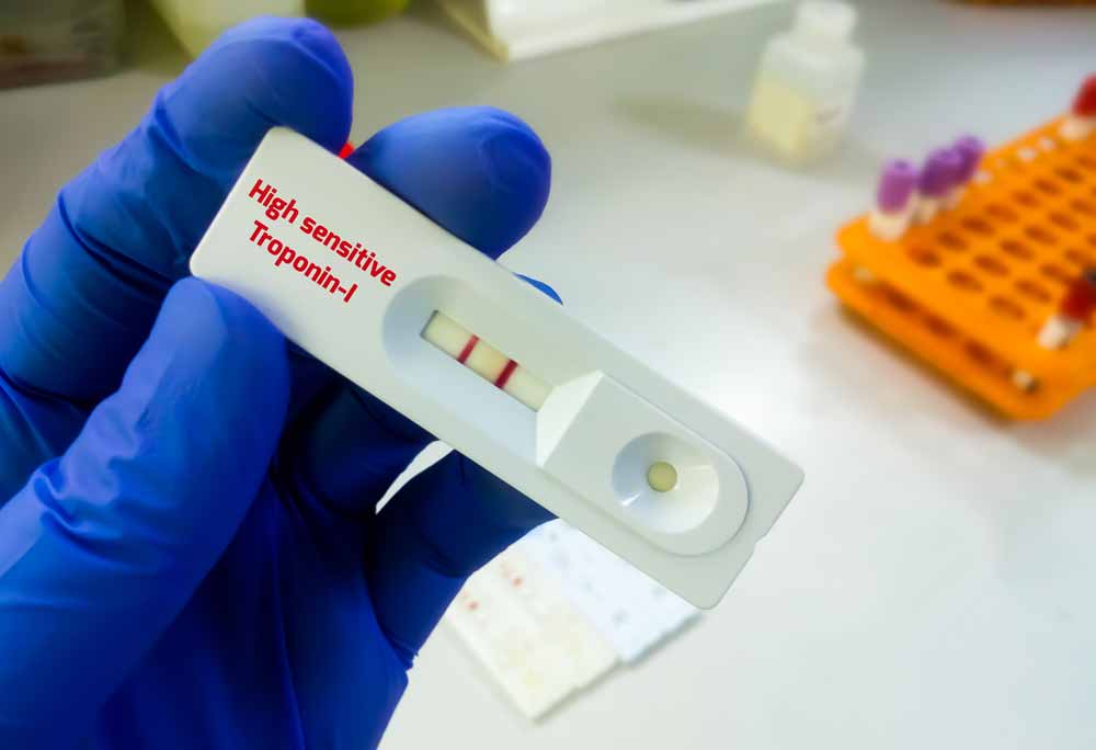
In Takotsubo syndrome, there is a moderate increase in troponin I/T and CK levels, which do not correlate with the significant impairment of regional myocardial contractility detected on echocardiography.
Blood analysis for natriuretic peptides
Elevated levels of BNP and NT-proBNP, markers of heart failure, are observed in Takotsubo syndrome patients due to cardiomyocyte overstretching from overload.
Electrocardiography
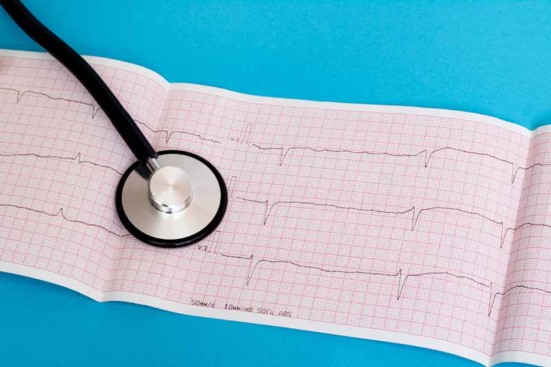
Characteristic ECG findings include ST segment elevation in precordial leads, elevation in AVR lead, diffuse T wave inversion, QT interval prolongation, and rarely, ST segment depression. Some experts highlight ST-segment elevation in leads V1-V3 along with AVR as characteristic of Takotsubo syndrome.
Transthoracic echocardiography
The characteristic echocardiographic signs of Takotsubo syndrome include transient systolic dysfunction of the left ventricle, transient akinesis of the apical segments of the myocardium of the left ventricle in combination with hyperkinesis of the basal part, apical dilatation of the myocardium of the left ventricle, sometimes obstruction of the outflow tract of the left ventricle.
Coronarography
Coronary angiography is important in the differential diagnosis of Takotsubo syndrome from acute myocardial infarction. In patients with Takotsubo syndrome, intact coronary arteries or coronary arteries without hemodynamically significant stenosis (stenosis <50%) are usually visualized. However, it should be noted that the presence of hemodynamically significant stenosis does not exclude the diagnosis of Takotsubo syndrome.
Ventriculography
Ventriculography allows us to evaluate the morphology and function of the left ventricle. Patients with Takotsubo syndrome are characterized by transient dysfunction and distension of the middle and apical segments of the left ventricle, due to which the heart takes on the shape of an octopus.
MRI of the heart
Characteristic MR signs of Takotsubo syndrome include transmural edema in areas of reduced local myocardial contractility, and absence of late gadolinium accumulation, while the basal parts of the left ventricle remain normal.
Diagnostic criteria
Among the existing diagnostic criteria for Takotsubo syndrome, the Mayo Clinic diagnostic criteria have the greatest practical significance :
- Transient dyskinesia of the middle segments of the left ventricle; Localized myocardial contractility impairment extending beyond the perfusion territory of a single epicardial artery
- Absence of obstructive coronary artery disease or angiographic evidence of acute plaque rupture
- New ECG changes (ST-segment elevation and/or T wave inversion) or a moderate elevation in troponin levels
- Absence of pheochromocytoma or myocarditis
In addition, you can use the International Takotsubo diagnostic criteria:
- Transient left ventricular dysfunction, commonly involving apical, mid-ventricular, basal, or focal dilatation; sometimes extending beyond the perfusion territory of a single epicardial artery; occasionally involves the right ventricle
- Emotional, physical, or combined triggers preceding symptom onset, though not always present
- Neurological disorders (e.g., subarachnoid hemorrhage, stroke/transient ischemic attack, convulsions) as potential triggers, including pheochromocytoma
- New ECG changes (ST-segment elevation/depression, T wave inversion, QT interval prolongation), occasionally normal ECG findings
- Moderate elevation in cardiac biomarkers (troponin, CPK) in most cases; significant elevation in BNP and NT-proBNP levels
- Severe coronary artery disease does not exclude Takotsubo syndrome diagnosis
- Absence of signs of infectious myocarditis
- Predominantly affects menopausal women
As well as the criteria of the Heart Failure Association and the European Community:
- Transient regional myocardial contractility disorder affecting both the left and right ventricles, often triggered by emotional or physical stress, although not always
- Involvement of myocardial areas beyond the perfusion territory of a single epicardial artery, often resulting in circular dysfunction of affected ventricular segments
- Absence of obstructive coronary artery disease, including acute plaque rupture, thrombus formation, coronary artery dissection, or other conditions that could explain transient left ventricular dysfunction (e.g., hypertrophic cardiomyopathy, viral myocarditis)
- New and reversible ECG changes during the acute phase (3 months), such as ST-segment elevation/depression, T wave inversion, QT interval prolongation, or complete left bundle branch block
- Significant elevation in B-type natriuretic peptide (BNP) and N-terminal pro-BNP (NT-proBNP) levels during the acute phase
- Moderate elevation in cardiac troponin levels
- Restoration of left ventricular systolic function confirmed by cardiac MRI within 3 to 6 months of onset
Differential diagnosis
In clinical practice, distinguishing Takotsubo syndrome from acute myocardial infarction is crucial.
Takotsubo Syndrome vs. Acute Myocardial Infarction:
Clinical Presentation:
- Both conditions present with acute chest pain and/or shortness of breath. However, Takotsubo syndrome often follows intense psycho-emotional stress, while acute myocardial infarction is associated with traditional cardiovascular risk factors (e.g., dyslipidemia, hypertension, diabetes).
ECG Findings:
- Takotsubo syndrome typically exhibits diffuse ST-segment elevation and/or T-wave inversion, whereas acute myocardial infarction shows ST-segment elevation and/or T-wave changes specific to the coronary artery territory affected.
Cardiac Biomarkers:
- In Takotsubo syndrome, troponin I/T levels are moderately elevated, with significantly elevated brain natriuretic peptide levels. Conversely, acute myocardial infarction is characterized by significantly elevated troponin I/T levels and moderately elevated brain natriuretic peptide levels.
Echocardiography:
- Both conditions demonstrate local myocardial contractility abnormalities and left ventricular systolic dysfunction on echocardiography. However, in Takotsubo syndrome, these changes are transient and involve regions beyond the territory of a single coronary artery, distinguishing it from acute myocardial infarction.
Coronary angiography
- Coronary angiography typically reveals intact arteries in Takotsubo syndrome and coronary artery obstruction in acute myocardial infarction. However, the absence of obstructive coronary lesions does not rule out myocardial infarction, and significant coronary stenosis does not exclude Takotsubo syndrome.
During ventriculography, systolic swelling of the segments of the left ventricle, most often apical, is determined.
Patients with Takotsubo syndrome show no late gadolinium on cardiac MRI, whereas subendocardial or transmural late gadolinium is present in acute myocardial infarction.
It is also worth mentioning the key differences between Takotsubo syndrome and other diseases that are accompanied by chest pain, including acute myocarditis, angina pectoris, pulmonary embolism, dissecting aortic aneurysm, and acute pericarditis.
Takotsubo Syndrome vs. Myocarditis:
Both conditions present with chest pain, shortness of breath, and elevated troponin levels, but in Takotsubo syndrome, troponin elevation is moderate and disproportionate to myocardial involvement. ECG changes may include ST segment and T wave abnormalities, with myocarditis often featuring conduction disturbances and arrhythmias.
- Takotsubo syndrome in 90% of cases develops in women over 50 years of age, while acute myocarditis most often affects young people without sexual dominance.
- Takotsubo syndrome in most cases is preceded by severe stress, while myocarditis is an infectious disease.
- Clinical symptoms such as chest pain, shortness of breath, and palpitations are characteristic of both Takotsubo syndrome and myocarditis.
- Blood troponin levels are elevated in both Takotsubo syndrome and myocarditis. However, in Takotsubo syndrome, this increase is insignificant and disproportionate to the area of the affected myocardium, and in myocarditis, troponin is usually significantly increased and correlates with the area of myocardial damage.
- The level of brain natriuretic peptide is significantly increased in Takotsubo syndrome and is often moderately increased in myocarditis.
- On the ECG of a patient with Takotsubo syndrome and myocarditis, changes in the ST segment and T wave can often be detected. Unlike Takotsubo syndrome, myocarditis is also characterized by conduction disturbances and arrhythmias.
- Echocardiography is important for differential diagnosis. Thus, in Takotsubo syndrome, swelling of the apical segments of the myocardium with intact basal sections is determined, which is not characteristic of myocarditis.
- Myocardial edema can often be detected on MRI of the heart in Takotsubo syndrome and myocarditis. However, in the acute stage of Takotsubo syndrome, unlike myocarditis, there is no late accumulation of gadolinium.
Takotsubo Syndrome vs. Angina Pectoris:
While both conditions exhibit chest pain, angina pain typically lasts up to 20 minutes and resolves with rest or nitrate use, whereas Takotsubo syndrome pain lasts longer and is nitrate-resistant. ECG findings in Takotsubo syndrome may show ST segment elevation and T-wave inversion, unlike angina.
- Both angina and Takotsubo syndrome are characterized by radiating chest pain. However, in angina, the pain lasts up to 20 minutes and disappears at rest or after taking nitrates, while in Takotsubo syndrome the pain lasts longer and is not relieved by nitrates.
- The ECG of a patient with angina is usually normal or shows ST-segment depression in the background of pain, whereas patients with Takotsubo syndrome often have ST-segment elevation and T-wave inversion.
- In patients with angina pectoris, the levels of markers of myocardial necrosis are normal, while those with Takotsubo syndrome are moderately elevated.
- A coronary angiogram of a patient with angina usually shows hemodynamically significant stenosis, while in Takotsubo syndrome the coronary arteries are either intact or with insignificant stenosis.
Takotsubo Syndrome vs. Pulmonary Embolism:
Both present with chest pain and shortness of breath, and troponin levels may be mildly elevated. ECG findings may include sinus tachycardia and T-wave inversion. However, pulmonary embolism often exhibits the classic S I Q III T III pattern, while Takotsubo syndrome typically shows ST segment elevation in precordial leads.
- Thromboembolism of the pulmonary artery and Takotsubo syndrome are characterized by complaints of anginal pain and shortness of breath.
- Blood troponin levels may be mildly elevated in both pulmonary embolism and Takotsubo syndrome.
- ECG in patients with pulmonary embolism and Takotsubo syndrome often shows sinus tachycardia and T-wave inversion. Pulmonary embolism is characterized by a classic S I Q III T III pattern, while Takotsubo syndrome is characterized by ST-segment elevation in the precordial leads.
- Echocardiography is important for differential diagnosis. Thus, in Takotsubo syndrome, ballooning of the apical segments of the myocardium of the left ventricle with intact basal sections is determined, while in thromboembolism of the pulmonary artery – akinesia or hypokinesia of the free wall of the right ventricle with preserved contractility of the apex of the right ventricle (McConnell’s sign), 60/60 sign, dilatation and dysfunction right ventricle, acute tricuspid regurgitation occurred.
Takotsubo Syndrome vs. Aortic Dissection:
Aortic dissection presents with sudden, severe chest pain that may radiate, often accompanied by asymmetric pulses and blood pressure. ECG may show ST segment elevation, but in different leads compared to Takotsubo syndrome. Imaging modalities such as echocardiography and chest X-ray can reveal aortic dilatation, distinguishing it from Takotsubo syndrome.
- Patients with a dissecting aortic aneurysm complain of sudden, unbearable, tearing pain in the chest, which radiates to the neck, back, and lower limbs, and is often migratory in nature. Although the pain in Takotsubo syndrome is also intense, however, not as much as in aortic dissection. During the physical examination of a patient with a dissecting aortic aneurysm, an asymmetry of the pulse and blood pressure on the extremities is revealed compared to the other side, in the case of involvement of the aortic valve in the process, a diastolic murmur over the aorta and a high pulse pressure are heard.
- On the ECG of a patient with a dissecting aortic aneurysm, as in a patient with Takotsubo syndrome, ST-segment elevation can often be detected, however, in Takotsubo syndrome, ST-segment elevation is visualized in the precordial leads, while in aortic dissection involving the right coronary artery, in leads II, III , AVF.
- Echocardiography and chest X-ray of have important diagnostic value. On echocardiography and chest X-ray, in the case of aortic arch dissection, dilatation of the affected part of the aorta is revealed, which is not the case with Takotsubo syndrome.
Takotsubo Syndrome vs. Acute Pericarditis:
Chest pain in acute pericarditis worsens with lying down and improves with sitting, and patients may have symptoms of infectious disease. ECG changes include diffuse ST-segment elevation. Echocardiography may reveal pericardial thickening and fluid accumulation, distinguishing it from Takotsubo syndrome.
- Acute pericarditis is characterized by chest pain that increases when lying down and decreases when sitting, which is not characteristic of Takotsubo syndrome. In addition, patients with acute pericarditis often have a history of symptoms of an infectious disease, including fever, catarrhal phenomena, and others, which are also not characteristic of Takotsubo syndrome.
- Clinical blood analysis of a patient with acute pericarditis reveals signs of an inflammatory process, which is not the case with Takotsubo syndrome.
- On the ECG of a patient with acute effusion pericarditis, as in a patient with Takotsubo syndrome, ST-segment elevation, and T-wave inversion can often be detected, however, in Takotsubo syndrome, these changes are visualized in the precordial leads, while in acute pericarditis, ST-segment elevation is diffuse.
- Echocardiography is important for differential diagnosis. Thus, in Takotsubo syndrome, ballooning of the apical segments of the myocardium of the left ventricle with intact basal sections is determined, while in pericarditis – thickening and compaction of the pericardial leaves, free fluid in the pericardium, signs of diastolic dysfunction.
Classification
Anatomical Variants of Takotsubo Syndrome:
- Apical or typical (75-80%)
- Mid-left ventricular (10-20%)
- Basal or inverted (5%)
- Biventricular (<0.5%)
- Focal (very rare)
Complication
The most frequent complications of Takotsubo syndrome include: acute heart failure (12-45%), left ventricular outflow tract obstruction (10-25%), mitral regurgitation (14-25%), and cardiogenic shock (6-20%).
Less common complications include: atrial fibrillation (5-15%), left ventricular thrombus (2-8%), sinus node arrest (4-6%), and AV block (5%).
Rare complications of Takotsubo syndrome include tachyarrhythmia (2-5%), bradyarrhythmia (2-5%), ventricular tachycardia of the pirouette type (2-5%), death (1-4.5%), ventricular tachycardia/fibrillation (3%), acute defect of the interventricular membrane (up to 1%).
| Most Frequent Complications: | Less Common Complications: | Rare Complications: |
| Acute heart failure (12-45%) Left ventricular outflow tract obstruction (10-25%) Mitral regurgitation (14-25%) Cardiogenic shock (6-20%) | Atrial fibrillation (5-15%) Left ventricular thrombus (2-8%) Sinus node arrest (4-6%) AV block (5%) | Tachyarrhythmia (2-5%) Bradyarrhythmia (2-5%) Ventricular tachycardia of the pirouette type (2-5%) Death (1-4.5%) Ventricular tachycardia/fibrillation (3%) Acute defect of the interventricular membrane (up to 1%) |
Treatment
Patients with Takotsubo syndrome require inpatient treatment, the main goal of which is to maintain hemodynamics until the systolic function of the left ventricle is restored and to prevent complications. As a rule, supportive treatment lasts several weeks.
Acute phase therapy
In the acute phase of Takotsubo syndrome, treatment aims to stabilize hemodynamics and prevent complications. Treatment strategies vary based on the presence of heart failure signs and the patient’s hemodynamic status.
Patients without heart failure symptoms typically receive beta-blockers, ACE inhibitors, or ARBs.
For patients exhibiting signs of lung congestion without hypotension, diuretics and nitrates are administered to reduce venous return to the heart. Additionally, ACE inhibitors, ARBs, or beta-blockers may be prescribed to alleviate congestion and reduce myocardial workload.
Patients with unstable hemodynamics require urgent echocardiography to assess for left ventricular outflow tract obstruction. Beta-blockers are indicated if obstruction is present, while inotropes are avoided.
Inotropic agents such as dopamine, dobutamine, milrinone, or levosimendan may be administered to patients without left ventricular outflow tract obstruction.
In severe cases, mechanical circulatory support like extracorporeal membrane oxygenation or intra-aortic balloon counterpulsation may be necessary.
Anticoagulant therapy is initiated and continued until left ventricular systolic function is restored, especially if there’s a significant decrease in left ventricular ejection fraction or a large area of akinesis/hypokinesis.
Treatment of complications
Management of acute heart failure and left ventricular outflow tract obstruction is crucial.
Ventricular arrhythmias are treated according to established guidelines, with beta-blockers or magnesium favored over drugs prolonging PQ interval (you should avoid the use of drugs that prolong the PQ interval). Electrical cardioversion may be necessary in some cases.
If left ventricular thrombi are detected, anticoagulation therapy is recommended for at least 3 months or until resolution.
Long-term therapy
According to the results of a retrospective analysis of large international registries, the positive effect of ACE inhibitors and BRAII in preventing the recurrence of Takotsubo syndrome was also demonstrated.
Experts’ opinions on the need for long-term beta-blockers to prevent relapses differ. On the one hand, long-term use of beta-blockers is pathogenetically determined, because it suppresses the activity of the sympathetic nervous system. On the other hand, according to observations, a third of patients who took beta-blockers for a long time developed a relapse.
Given the high frequency of mental health disorders among patients with Takotsubo syndrome, such as depression, anxiety disorders, and substance abuse, correction of these disorders is important to prevent the recurrence of Takotsubo syndrome.
To assess the level of anxiety, use the Hamilton scale:
Register on our website right now to have access to more learning materials!
Subscribe to our pages:
Acute Pulmonary Edema: Emergency Care Algorithm – Should We Remove or Redistribute the Fluid?
Case Presentation: A 64-year-old man was transported to the emergency department by ambulance due to…
Сounseling a patient with suspected Takotsubo-syndrome OSCE guides
The onset of the consultation Wash hands and put on PPE if necessary. Introduce yourself…
Counseling of a patient with symptomatic bradycardia – OSCE guide
https://clincasequest.hospital/course/interrupted-symphony/ The onset of the consultation Wash hands and put on PPE if necessary. Introduce…
Symptomatic bradycardia
Symptomatic bradycardia occurs when the heart rate drops below 50 beats per minute. Most often,…
Baseline Cardiovascular Risk Assessment in Cancer Patients Scheduled to Receive Cardiotoxic Cancer Therapies (Anthracycline Chemotherapy) – Online Calculator
Baseline cardiovascular risk assessment in cancer patients scheduled to receive cardiotoxic cancer therapies (Anthracycline Chemotherapy)…
SAVED VTE Score
SAVED score for venous thromboembolism risk stratification in patients with multiple myeloma receiving immunomodulators. [ezfc…
Resources
- Takotsubo Syndrome: Pathophysiology, Emerging Concepts, and Clinical Implications. Trisha Singh, Hilal Khan, David T. Gamble et al. Circulation. 2022; 145:1002-1019. DOI: 10.1161/CIRCULATIONAHA.121.055854
- International Expert Consensus Document on Takotsubo Syndrome (Part II): Diagnostic Workup, Outcome, and Management. Jelena-Rima Ghadri, Ilan Shor Wittstein, Abhiram Prasad et al. European Heart Journal. 2018; 39: 2047–2062. doi:10.1093/eurheartj/ehy077
- Stress Cardiomyopathy Diagnosis and Treatment. Horacio Medina de Chazal, Marco Giuseppe Del Buono, Lori Keyser-Marcus et al. Journal of the American College of Cardiology. 2018; 72 (16): 1955. doi.org/10.1016/j.jacc.2018.07.072
- Takotsubo syndrome: unraveling the enigma of the broken heart syndrome?-a narrative review. Cardiovascular Diagnosis and Therapy. 2023;13(6):1080-1103. https://dx.doi.org/10.21037/cdt-23-283.

