
Chest CT Scan
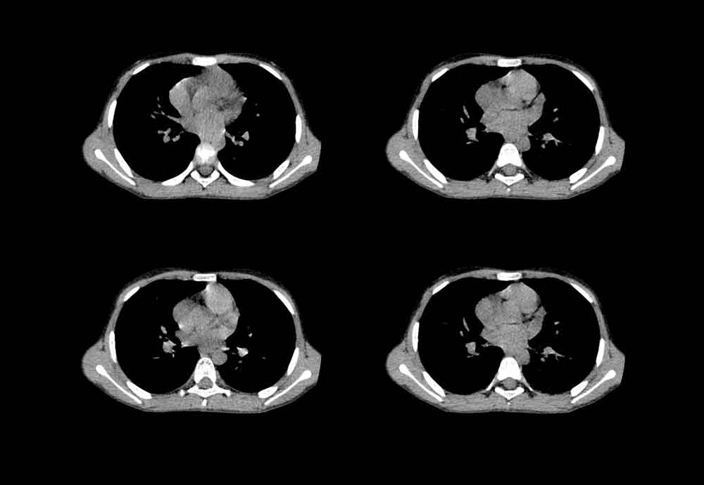
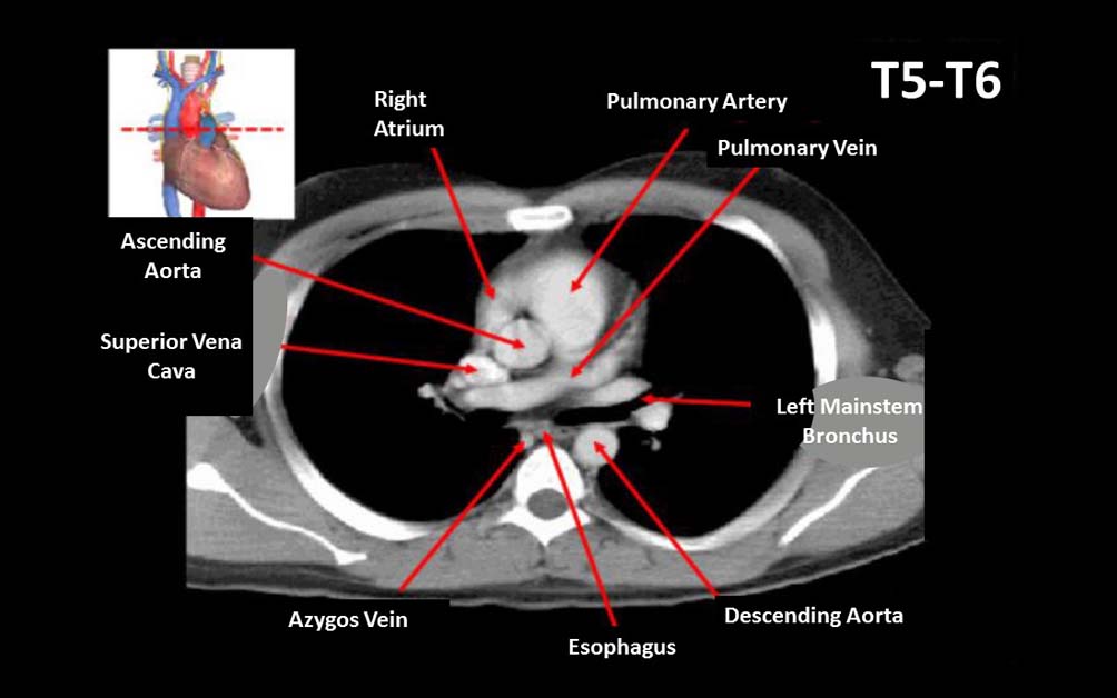
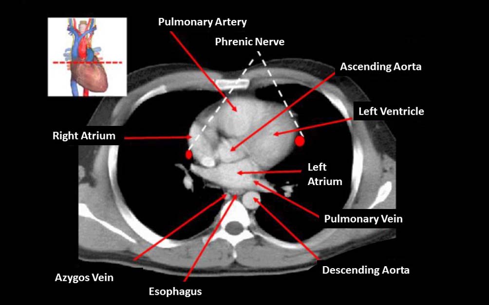
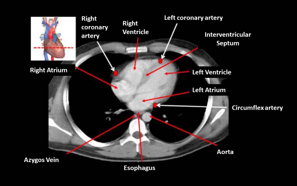
To analyze a CT scan of the chest, imagine that the patient is lying on his back and you are at the foot of the patient’s bed.
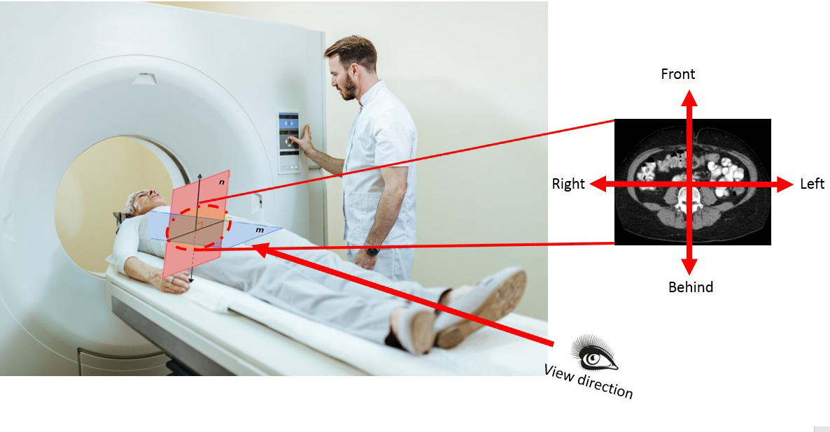
In order to identify the structures of the chest, it is important to determine the orientation (right, left parts of the image, anterior and dorsal – posterior structures).
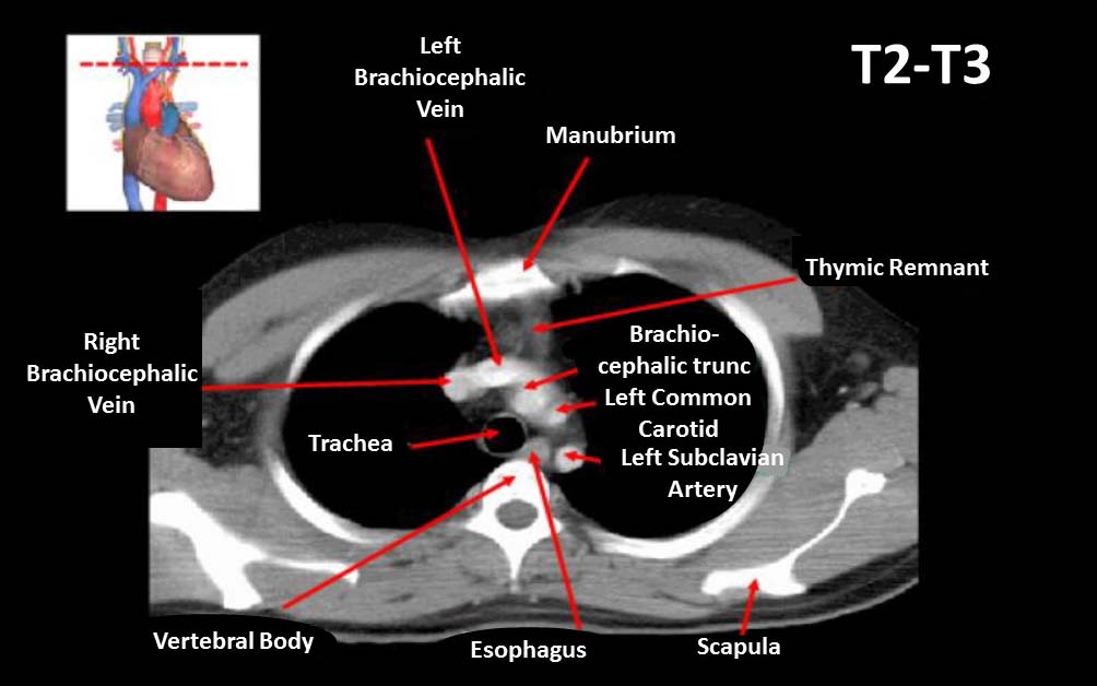
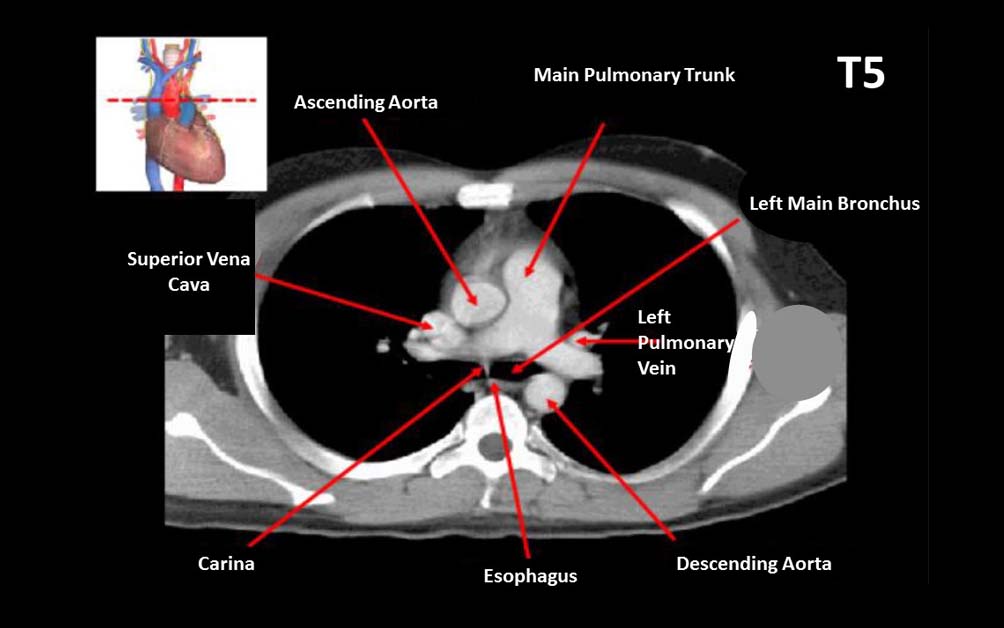
Depending on the location (upper mediastinum, lower mediastinum), the presence of certain structures of the mediastinum will be determined.
The border between the upper and lower mediastinum passes at the height of the IV thoracic vertebra dorosally and ventrally – the Lewis angle (between the manubrium and the body of the sternum).

Register on our website right now to have access to more learning materials!
Subscribe to our pages:

Attempt-Based Analysis: A New Paradigm for Assessing Clinical Reasoning in Simulation
ClinCaseQuest is proud to highlight the presentation of Tetiana Antofiichuk, MD, PhD, at the international…

ClinCaseQuest presented the first experience of analyzing branching simulation logs based on artificial intelligence at the “Medical Simulation: A Look into the Future” Conference
ClinCaseQuest is pleased to share our participation in the scientific and practical conference “Medical Simulation:…

AI-Driven Patient Simulator – Quick Feedback
🗣️ We Value Your Feedback Thank you for your interest in the AI-Driven Patient Simulator!…
Get Early Access AI-Driven Patient Simulator
Try the AI-Driven Patient Simulator and explore the future of medical education 🧠 Try the…

Celebrating a Major Milestone: SESAM Recognizes ClinCaseQuest’s Defragmented Debriefing Model as an Advancement in Clinical Simulation 2024
At ClinCaseQuest, we are thrilled to announce an outstanding achievement in the field of medical…

Acute Pulmonary Edema: Emergency Care Algorithm – Should We Remove or Redistribute the Fluid?
Case Presentation: A 64-year-old man was transported to the emergency department by ambulance due to…