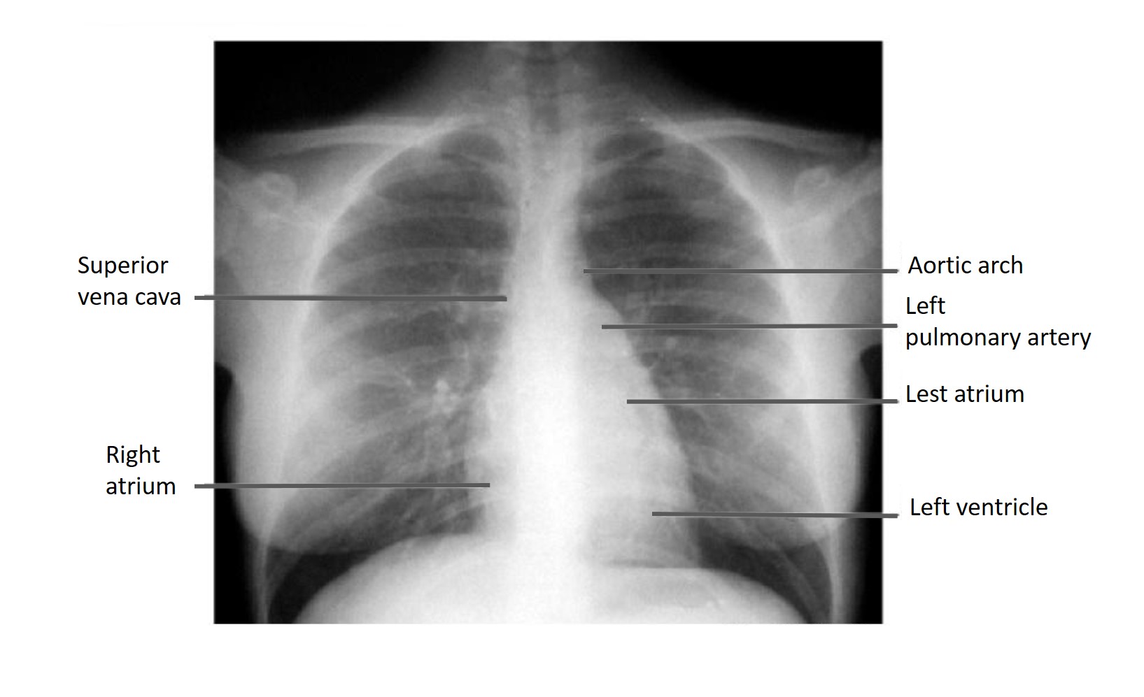
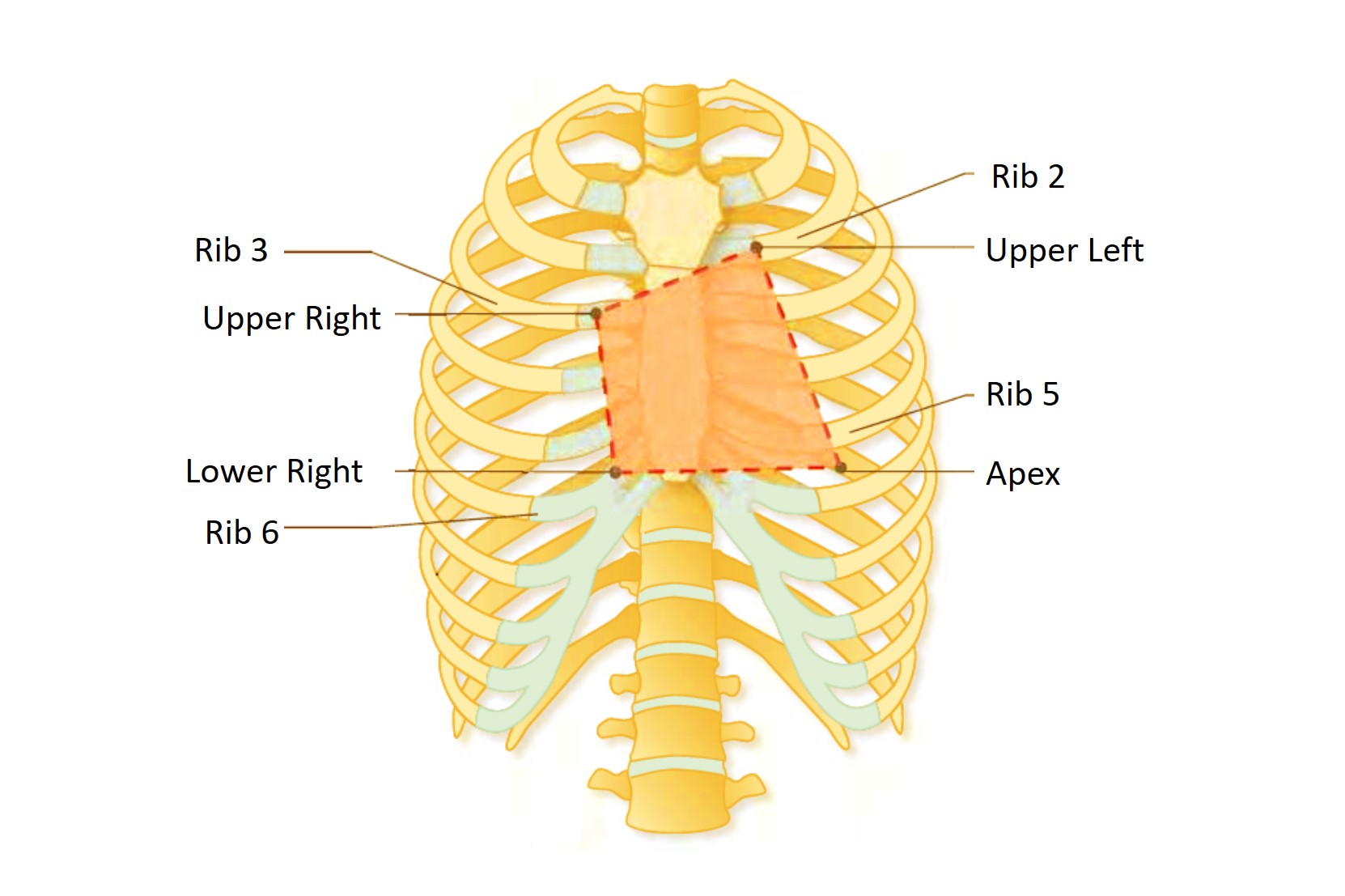
X-ray Heart Borders
According to the radiograph of the chest, the boundaries of the heart are formed:
- The right border of the heart is the superior vena cava, the right atrium. The anterior wall of the heart is the right ventricle.
- Left border of the heart – aortic arch, left pulmonary artery, left atrium, left ventricle
- The lower border of the heart is the left ventricle.
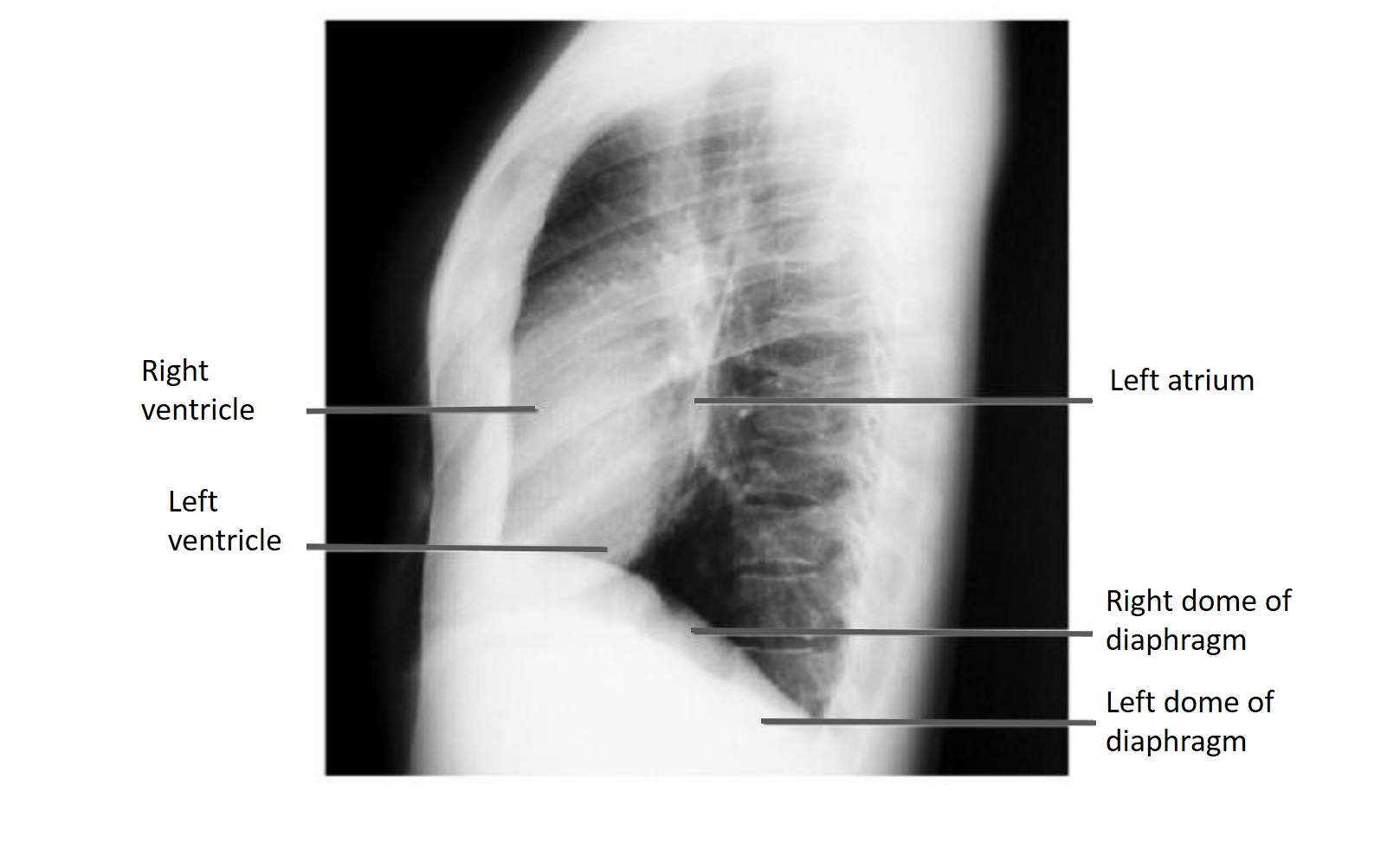
- The right border extends between the margin of the third right costal cartilage to the sixth right costal cartilage just to the right of the sternum.
- The left border extends between the fifth left intercostal space to the second left costal cartilage.
- The inferior border extends from the sixth right costal cartilage to the fifth left intercostal space at the midclavicular line.
- The superior border extends from the inferior margin of the second left costal cartilage to the superior margin of the third costal cartilage.
Register on our website right now to have access to more learning materials!
Subscribe to our pages:

Baseline Cardiovascular Risk Assessment in Cancer Patients Scheduled to Receive Cardiotoxic Cancer Therapies (Anthracycline Chemotherapy) – Online Calculator
Baseline cardiovascular risk assessment in cancer patients scheduled to receive cardiotoxic cancer therapies (Anthracycline Chemotherapy)…

SAVED VTE Score
SAVED score for venous thromboembolism risk stratification in patients with multiple myeloma receiving immunomodulators.
Loading...